
Radiographic Interpretation: Getting the Most out of Dental Radiographs.
Presented by Ms. Marlena Shore
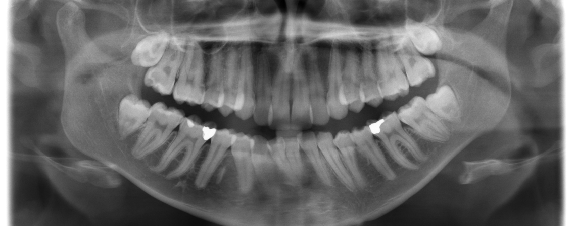
Description
Radiology is a tool used in Dentistry every day to provide the clinician important information that cannot be seen in a clinical exam. Intraoral and extraoral radiographs aide in assessments, periodontal therapy and restorative treatment.
These images allow for the view of calculus deposits, alveolar bone loss, faulty restorations, dental caries and pulpal changes. It is essential that dental professionals are able to acquire images that are diagnostically acceptable, while limiting the amount of radiation exposure by following the recommendations of ALARA. This course is intended to review normal anatomical landmarks in both the full mouth series and panoramic images.
By utilizing the techniques that will be discussed in this course, the clinician will be able to increase the accuracy of radiographic images and make a more valuable contribution to the dental practice.
LEARNING OBJECTIVES
- Identify normal anatomical landmarks, including radiolucencies and radiopacities on periapical, bitewing and panoramic radiographs.
- Identify carious lesions, periapical lesions and periodontal involvement radiographically.
- Understand supplementary techniques to use in the interpretation of both analog and digital radiographs.
Ms. Marlena Shore

MARLENA SHORE, RDHAP, MS has been a Registered Dental Hygienist since 1999, graduating from West Los Angeles College with Honors. In 2003 she continued her education and graduated in the first class from WLAC to become a Registered Dental Hygienist in Alternative Practice. …
Read MoreNo upcoming courses
Hands-On Workshop Courses

Dr. Pascal Magne
An Update on Aesthetic Anterior & Posterior Bonded Restorations: Mastering Direct and Indirect Techniques
Read More
Dr. Pascal Magne
PREDICTABLE “START TO FINISH” BIOMIMETIC DENTISTRY IN THE ANTERIOR DENTITION: From chemical treatments, to fragment reattachment, direct, semi-(in)direct and indirect restorations.
Read More