UPDATED PROGRAM - FROM MACRO TO MICRO - MICROVISION: TWO DAY HANDS ON COURSE
Register Now
Course Description
Introduction
In this course we will discuss all the steps of treating a patient, starting with photo video documentation, treatment planning, and finishing with bonding procedures. A complete protocol of tooth preparation for veneers and full crowns will be highlighted utilizing modified enamel chisels and the dental microscope. Multimedia will be utilised for optimal visual learning by using magnification photography and videos to focus on specific details required to improve your dental skills. We will focus on integration microscopic approach into a full mouth rehabilitation cases. The important role of magnification in dentistry will be further discussed.
DAY 1 - 9th JULY 2016
Theory
• MicroVision approach and philosophy
• Patient analysis and CR position detection
• Photography & video documentation
• Treatment planning, DSD, and presentation for the patient
• Ergonomics and the use of magnification, loupes and microscopes in prosthetics
• Co-operation with assistant
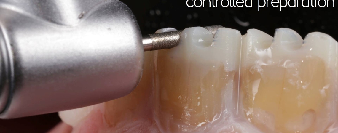
• Step by Step full crown preparation protocol
Hands On Course (Full crowns)
• Preparation of distal tooth for a full crown
• Impression taking techniques
• Bur choice
• Clamp choice
• Split dam
• Additional isolation: flosses, Teflon, retractive cords, silicon
• Step by Step preparation for full crowns
• Fabrication of provisional restorations
• Finishing and polishing
DAY 2- 10th JULY 2016
Theory
• Full mouth microvision rehabilation with veneers and onlays
• Planning and occlusion
• Communcation with the lab for optimal results
• Mockup transfer
• Mockup preparation
• Fabrication of provisional restorations - direct technique
• Impression
• Choice of ceramic materials
• Steps in the lab dentists should know
Hands On Course (Veneers)
• Ceramic veneer selection and protocols
• Veneer design preparations and material selection
• Step by Step procedure with MicroVision kit
• Isolation (rubber dam)
• Bur Choice
• Bonding protocols from A - Z
• Finishing and polishing
Hands-On Workshop Courses

Dr. Jeffrey Okeson
What Every Dentist Needs to Know About Temporomandibular Disorders (The Facts and The Fantasies) and A Participation Program
Read More
Dr Maxim Belograd
MASTERING TOOTH PREPARATION & BONDING OF CERAMIC RESTORATIONS - Crowns, Veneers, Onlays, Rubberdam, Cementation and Photography
Read More